Someone is looking at a photograph. We cannot see the photograph. Instead, we measure that person's brain activity. Then an image resembling what they saw appears on a computer screen.
It sounds like something from science fiction, but it is already possible in research laboratories.
More recently, AI pretrained on other people's data has been helping researchers obtain results faster.
The era in which AI redraws what the brain has seen has begun.
How closely do images drawn from brain signals resemble the originals?
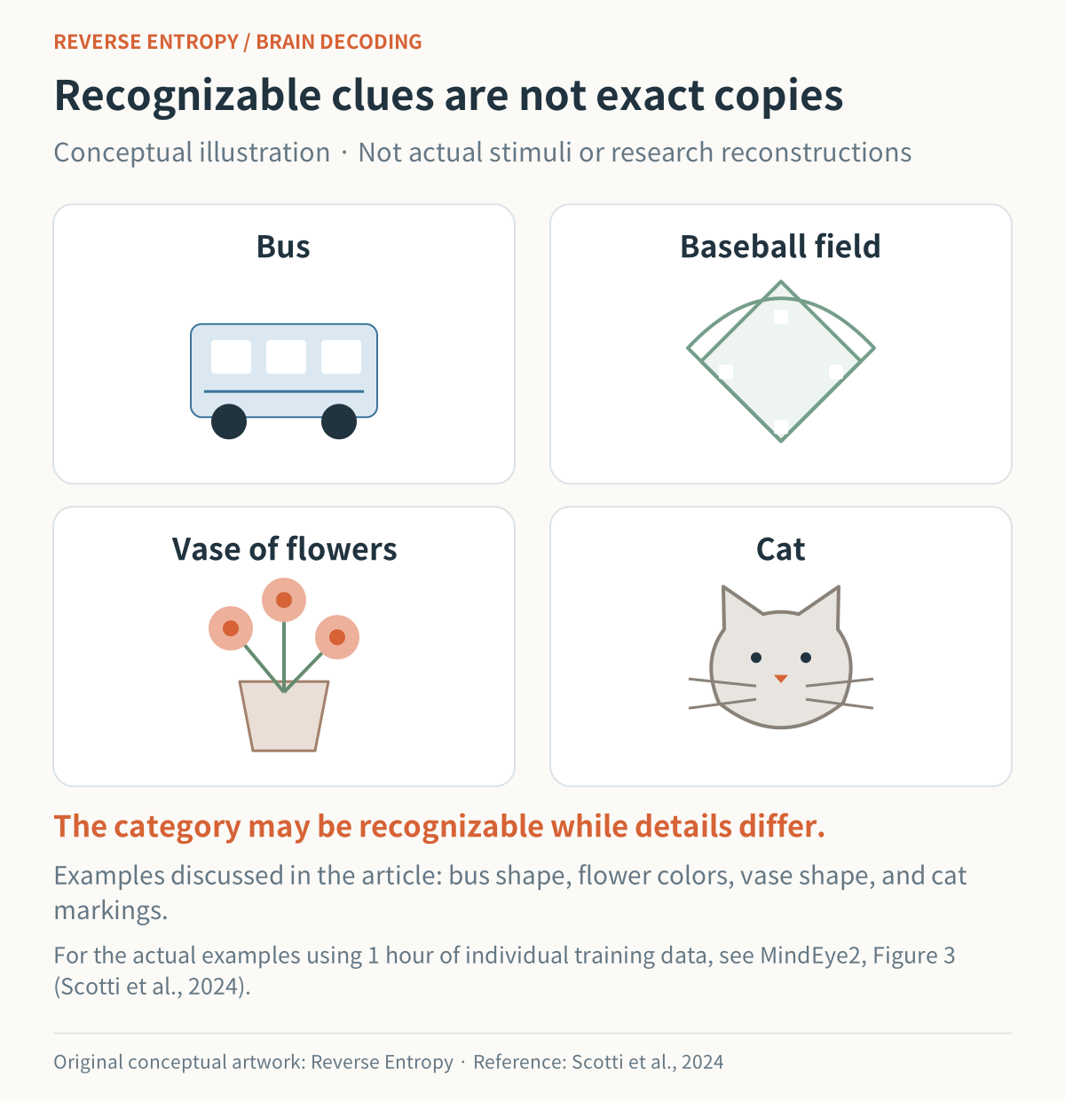
New conceptual illustration: Reverse Entropy. Research discussed: Scotti PS et al., ICML/PMLR, 2024, Figure 3. Original paper and research images · Paper license (CC BY 4.0). The original paper’s stimulus photographs are not reproduced here.
A bus, a baseball field, a vase of flowers, a cat. Figure 3 of the original MindEye2 paper published in 2024 contains actual examples of these scenes. I think we can certainly say those original examples look similar to this extent.
Of course, some differences are visible too.
The red double-decker bus became a differently shaped bus, and the colors of the flowers and the shape of the vase also changed.
The system captured substantial clues about the scene, but it did not copy the photograph exactly.
What is being measured inside the head?
The secret of this technology is to image the brain with MRI.
As people are shown photographs, researchers record brain responses that differ from scene to scene.
When a new brain signal arrives, the system compares it with the earlier records to infer what the image might be.
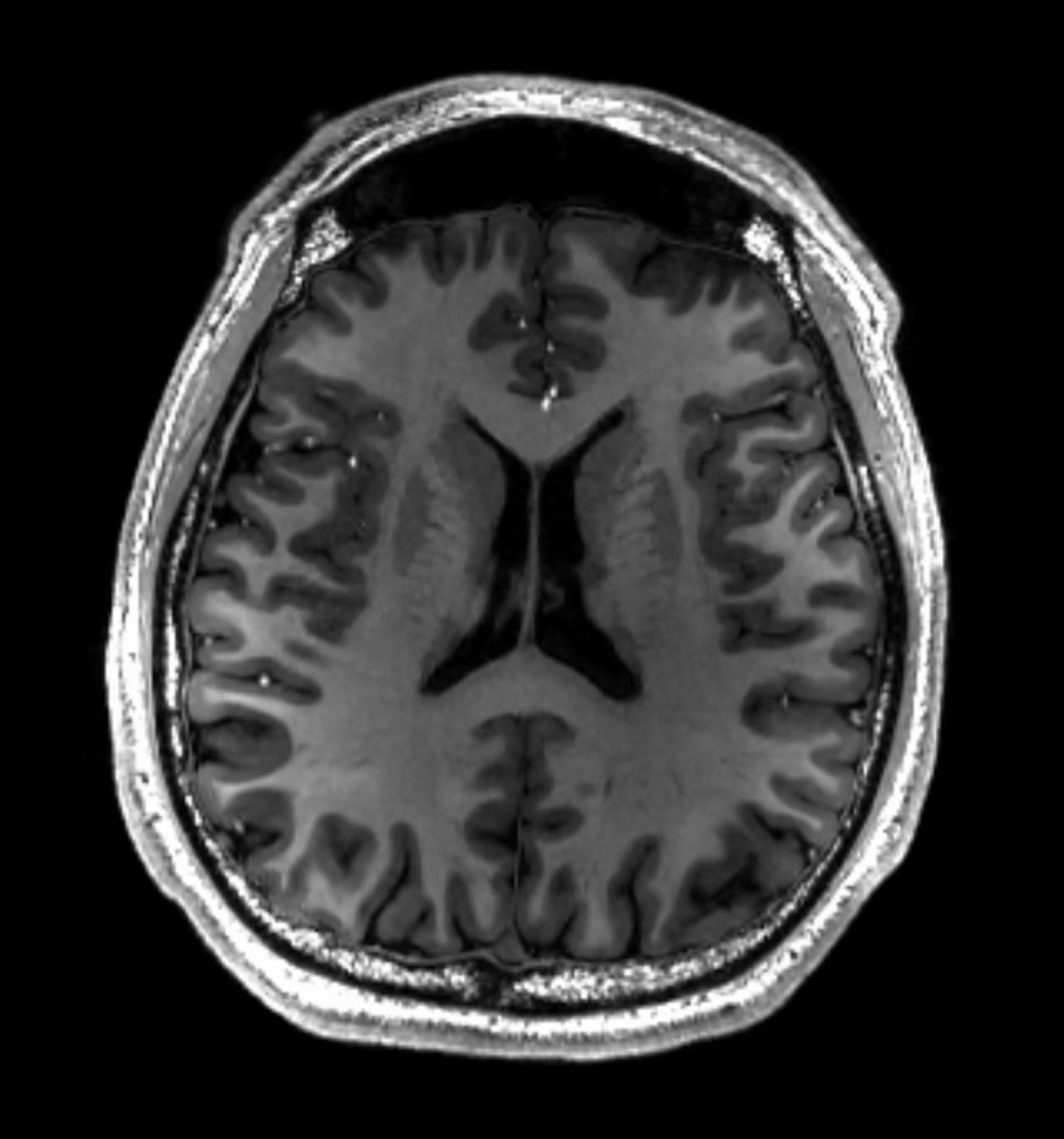
Source: Asnaebsa, Wikimedia Commons, 2022. Original image reproduced unchanged. Image source · License: CC BY-SA 4.0.
This is not the kind of MRI we commonly encounter in a hospital. The technique used in the representative studies is functional magnetic resonance imaging, or fMRI.
It indirectly reads brain responses through changes in blood oxygenation associated with neural activity.
This is different from taking a single conventional MRI image of the brain.
AI-assisted image · Learn more
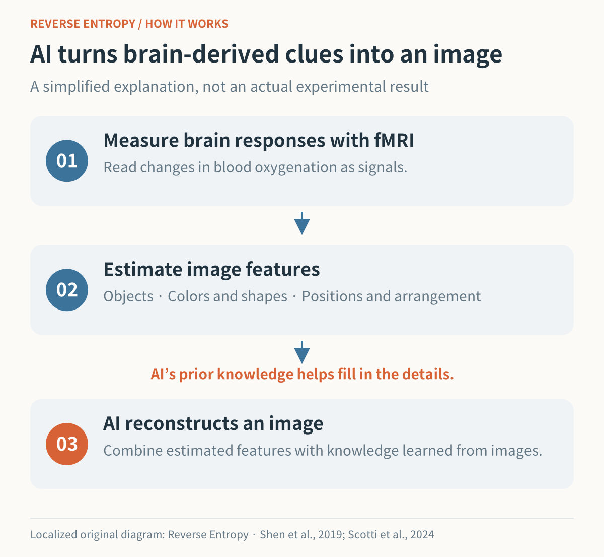
Source: Reverse Entropy’s original explanatory diagram, translated and redrawn for this edition. Based on Shen et al., 2019; Scotti et al., 2024. Research reference. This is a conceptual diagram, not an experimental result.
This is where generative AI comes in.
AI creates an image based on features inferred from the brain signals.
A technique for estimating the shapes and arrangement of objects is combined with a technique for expressing those clues as a natural-looking image.
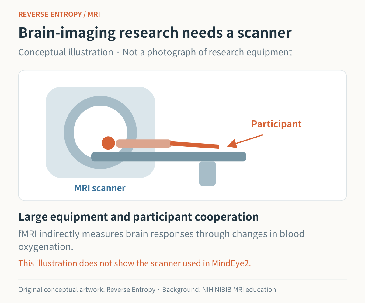
New conceptual illustration: Reverse Entropy. Background reference: NIH NIBIB, Magnetic Resonance Imaging educational material. The photograph used in the Korean article is not reproduced here.
Can it also reveal scenes that are not in front of us?
The next step is imagination: reading a scene someone is picturing mentally, rather than a photograph they are actually seeing.
The MIRAGE study, published in May 2026, reconstructed images from brain signals recorded while participants imagined specified images or concepts. The model was trained on data collected while people actually viewed photographs. [3]
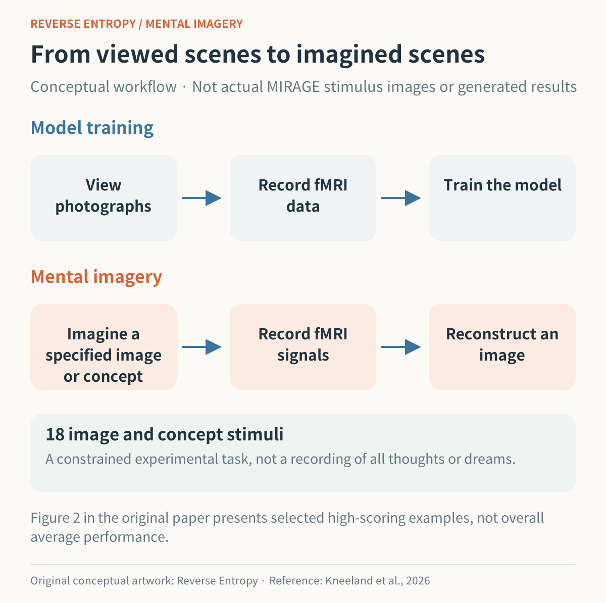
New conceptual workflow: Reverse Entropy. Research discussed: Kneeland R et al., PLOS Computational Biology, 2026, Figure 2. Original paper and research images · Paper license (CC BY 4.0). The original paper’s stimulus photographs and generated examples are not reproduced here.
In the original MIRAGE paper’s examples, clues such as a surfing pose or a bird are recognizable, but the colors and details differ from the originals.
The experiment was a constrained task involving 18 image and concept stimuli.
It did not record every freely arising thought, or film the dreams someone has throughout the night.
Interestingly, the model that best reconstructed images people were actually seeing was not necessarily the best at reconstructing imagined images.
Signals during imagination were weaker and less stable, requiring a decoding approach suited to them.
Suppose, for example, that a brain signal provides the clue “a dog on grass.” Generative AI can draw the texture of the fur, a glint in the eyes, and shadows cast by blades of grass.
But was every detail read from the brain? Some parts are filled in using what the AI has learned about dogs and grass.
The ability to produce a sharp, photograph-like picture is different from the ability to accurately reconstruct the scene someone actually saw.
“Spurious reconstruction from brain activity,” published in 2025, highlighted this problem in some image-reconstruction approaches. They produced plausible results for familiar kinds of images, but performance dropped substantially on a separate dataset whose categories did not overlap with those used in training.
When viewing a result, we therefore need to go a step beyond “Wow, it looks like a photograph.”
We need to check whether the number and positions of objects, their colors, and their relationships are correct too.
What excites me more is the potential for communication
If this technology becomes more accurate and convenient, it could help people who have difficulty expressing themselves through speech or movement communicate images in their minds. Researchers also identify communication assistance and research into consciousness and cognition as possible long-term applications.
From a physician's perspective, the possibility of developing a tool that lets patients convey visual experiences they find difficult to explain in words is also intriguing.
Scenes we have seen or imagined are becoming images we can show to someone else.
The goal ahead will not simply be how impressive a picture we can create. What matters will be how accurately that picture can capture the person's experience.
Research consulted
1. Scotti PS et al. MindEye2: Shared-Subject Models Enable fMRI-To-Image With 1 Hour of Data. ICML / PMLR 235:44038–44059 (2024).
2. Shen G, Horikawa T, Majima K, Kamitani Y. Deep image reconstruction from human brain activity. PLOS Computational Biology 15(1):e1006633 (2019).
3. Kneeland R et al. MIRAGE: Robust multi-modal architectures translate fMRI-to-image models from vision to mental imagery. PLOS Computational Biology 22(5):e1014263 (2026).
4. Shirakawa K et al. Spurious reconstruction from brain activity. Neural Networks 190:107515 (2025).
#Neuroscience #BrainScanning #fMRI #GenerativeAI #MindEye #BrainImaging #ArtificialIntelligence #ReverseEntropy