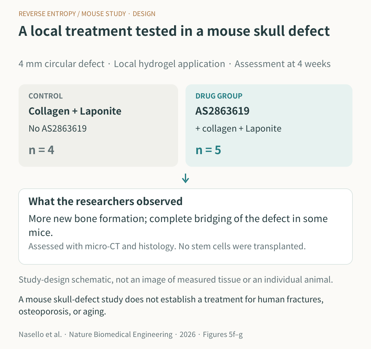
A hole is made in a mouse's skull.
When a gel containing a specific drug was applied to the site, new bone formed after four weeks, and in some cases bridging across the defect was observed.
What is interesting is that no stem cells were transplanted as part of this treatment.
The researchers were trying to influence the immune cells at the injury site.
This study was published in Nature Biomedical Engineering on October 23, 2026, by researchers at Harvard University and collaborating institutions. It is still a mouse bone-defect experiment, not a treatment result in humans, but it gives us something to think about regarding how tissues can be regenerated. Original study
The original Korean article included an AI-use disclosure for its explanatory artwork. Original disclosure information
Could changing the immune environment at an injury site help us make better use of the body's remaining capacity for regeneration?
Immune cells fight bacteria and viruses.
They also gather at sites of tissue injury, clear away damaged tissue, and send signals needed for recovery.
Early inflammation is therefore part of the wound-healing process. Problems arise when inflammation is too intense or lasts too long.
If the response to damage continues, the environment can become less favorable for new tissue to establish itself.
The cells this study focused on were regulatory T cells, or Tregs.
These cells keep other immune cells from overreacting and can be thought of as the immune system's brakes.
In damaged tissue, this regulatory function also influences recovery.
The researchers started with this question: could inducing regulatory T-cell activity at the injury site also help bone regenerate?
This does not mean that immune cells turn directly into bone cells. It is a strategy for adjusting the environment around the cells that build bone. Likewise, “without adding stem cells” in the title means that stem cells were not transplanted from outside the body. It does not mean that the body's own stem cells no longer have a role.
Keeping the drug at the injury site for longer
One way to use regulatory T cells therapeutically is to grow the cells outside the body and then administer them.
Alongside experiments delivering regulatory T cells locally, this research team investigated whether a drug could replace the process of growing and transplanting cells.
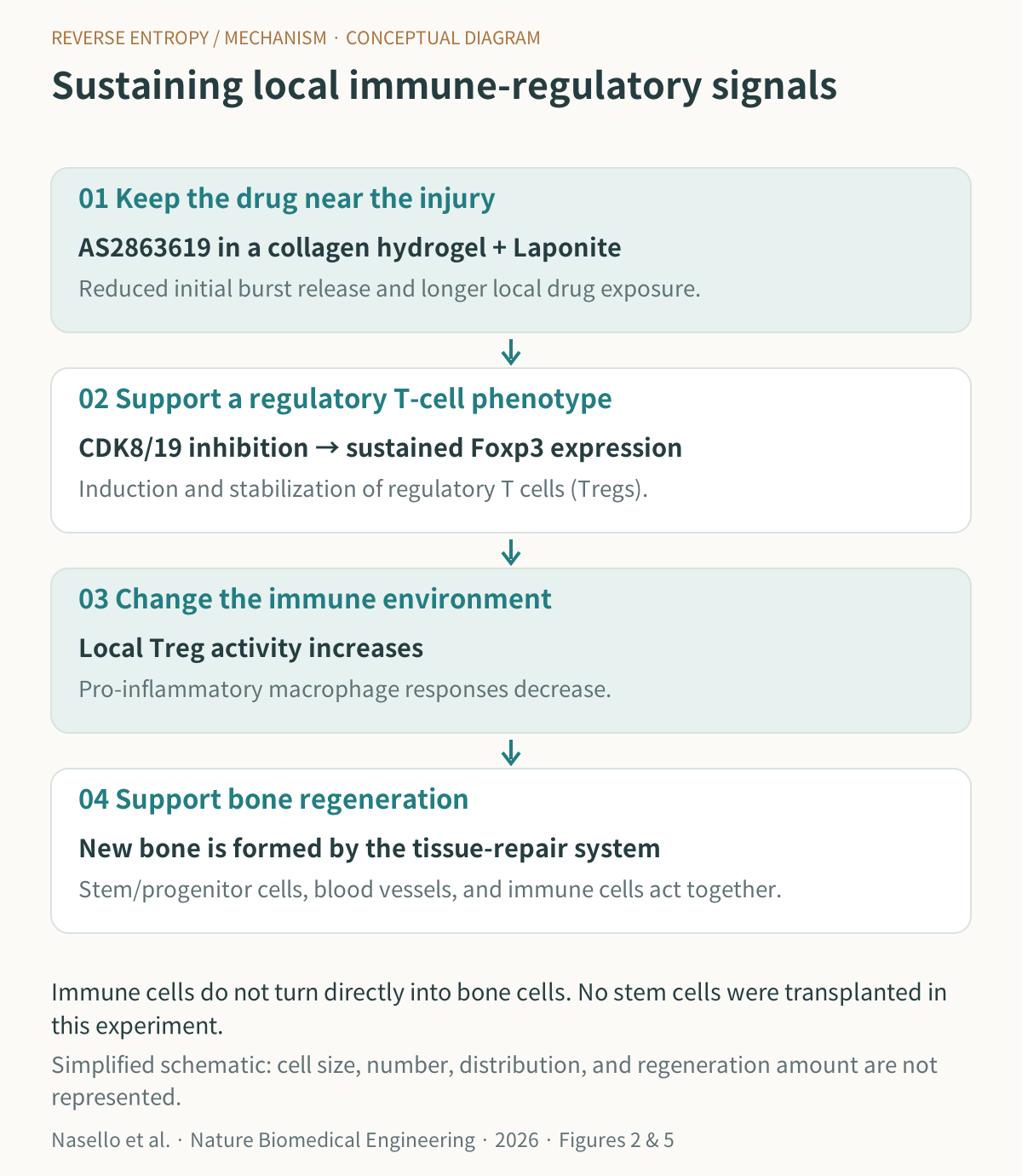
The compound they used was AS2863619. It inhibits the enzymes CDK8/19, inducing T cells to acquire regulatory T-cell characteristics.
A transcription factor called Foxp3 is important in this process. It regulates which genes a cell uses and is involved in regulatory T-cell differentiation and function.
Earlier research on CDK8/19 inhibition
In the team's cell-culture experiments, Foxp3 expression was maintained more stably in cells exposed to the drug for longer.
The duration of drug exposure therefore mattered.
To achieve this, the drug needed to be prevented from leaving the injury site all at once.
The researchers loaded it into a collagen hydrogel, a soft material that holds a large amount of water.
They went a step beyond simply mixing the drug into a gel.
They added nanoparticles called Laponite to reduce the initial burst of release and retain the drug for longer.
A signal needed for regeneration that appears briefly and then disappears can have a very different outcome from one maintained for a period of time.
A distinctive feature of this study is that the researchers designed the immune-modulating drug strategy and the material delivering the drug together.
Source: Created by Reverse Entropy from the publicly available abstract and Figures 2 and 5 of Nasello et al., Nature Biomedical Engineering, 2026. Image source Original graphic by Reverse Entropy.
What had changed in the mice's bones after four weeks?
The researchers created circular defects 4 mm in diameter in the skulls of mice.
Defects of this size do not readily fill in on their own.
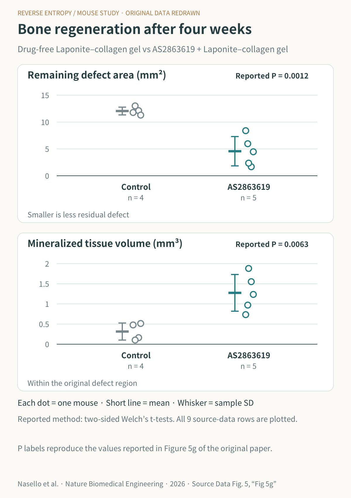
When they compared a drug-free hydrogel with a drug-loaded hydrogel, the proportion of regulatory T cells increased at the injury site, while pro-inflammatory macrophage responses decreased. Bone formation assessed by micro-CT also improved.
In the experiment with added Laponite, they compared a drug-free Laponite–collagen gel with the same material containing the drug.
Source: Nasello et al., Nature Biomedical Engineering, 2026, Source Data Fig. 5. New chart drawn by Reverse Entropy from the numerical data; the original copyrighted panel is not reproduced. Original paper figure
In some mice, new bone was reported to bridge the defect completely.
The significance lies in going beyond an experiment that changed only gene levels associated with bone formation: actual tissue formation was confirmed through imaging and histological examination.
Figure 5 and the experimental description
Can we also connect this with skin regeneration?
Reading this study brought to mind the process through which skin recovers after a stimulus.
In skin treatments, the environment in which tissue recovers afterward matters alongside the type of stimulus applied.
Skin recovery involves immune cells as well as fibroblasts, keratinocytes, and the cells that make up blood vessels.
Indeed, in a mouse skin-wound study, removing regulatory T cells delayed wound closure and epidermal recovery.
However, we cannot interpret this bone-regeneration experiment as showing improved recovery after laser or radiofrequency procedures in humans.
The tissues, types of injury, and treatment goals are different.
What interests me is the question for future research.
Alongside methods that stimulate collagen production, could we also make the conditions for appropriately regulated inflammation and tissue recovery after injury a target of treatment?
In anti-aging, I look at the conditions that make recovery possible
These findings do not immediately amount to a treatment for fractures or osteoporosis.
A localized defect in a mouse skull differs from a fracture in a weight-bearing bone, and from osteoporosis, which involves changes in bone metabolism throughout the body.
Nor did this study test whether it restored the recovery capacity of aged tissue or slowed aging itself.
Regulatory T cells are also not cells for which more is always better.
Because they suppress immune responses, developing this approach into a therapy will require determining how long they should act at the intended site and how the approach affects other immune functions, including protection against infection.
Even so, I think that when we consider anti-aging, we should look both at preserving healthy cells and tissues for longer and at maintaining the ability to recover when injury occurs.
Tissue regenerative capacity is not determined solely by the number of stem cells.
Blood vessels, the extracellular matrix, metabolic state, and signals from surrounding cells all act together.
This study showed that modulating one part of that system, the immune environment, can lead to actual bone regeneration.
Alongside technologies that supply new cells, technologies that create the conditions for cells already present to carry out repair may also advance.
What I would like to see next is whether this approach can produce adequate recovery in aged or diseased tissues as well.
References
1. Nasello G, Lafuente-Gómez N, Krajnovic L, et al. Hydrogel for sustained and localized delivery of a regulatory T cell-inducing agent promotes bone regeneration. Nature Biomedical Engineering. Published online October 23, 2026. DOI: 10.1038/s41551-026-01817-1
2. Akamatsu M, et al. Conversion of antigen-specific effector/memory T cells into Foxp3-expressing Treg cells by inhibition of CDK8/19. Science Immunology. 2019;4:eaaw2707. DOI: 10.1126/sciimmunol.aaw2707
3. Nosbaum A, et al. Cutting Edge: Regulatory T Cells Facilitate Cutaneous Wound Healing. Journal of Immunology. 2016;196:2010–2014. DOI: 10.4049/jimmunol.1502139
Gabriele Nasello and David J. Mooney, authors of the Nasello study, disclosed that they were named as inventors on a U.S. provisional patent application based on this research.