“I had under-eye filler years ago. Now, when I feel run-down or as if I’m coming down with something, I get sausage-like swellings under my eyes.”
I hear this from people who previously had filler in the tear troughs or upper cheeks. It looked natural at first, then swelling began to come and go. One day everything looks fine; the next, the area feels heavy and raised. After a few episodes, it is natural to be wary of further treatment and wonder whether filler simply disagrees with you.
I look at how much filler remains, the state of the surrounding tissues and what happens when swelling starts. Understanding the cause helps address the existing problem and plan any later treatment. My main principle is to handle support for deep hollows and care for the thin overlying skin separately.

1. First, find out why the area is swollen
Under-eye skin is thin; fat, muscle and ligaments sit close together, and lymphatic drainage matters. The same amount of filler can make contour changes and swelling more obvious here than elsewhere on the face.
The boundaries between under-eye and cheek tissues, including the malar septum, influence where fluid collects. Hydrophilic filler and impeded fluid drainage may together leave persistent puffiness. Anatomy study · Clinical report on malar edema
There is also a delayed inflammatory reaction (DIR): an area that was quiet for some time can develop swelling, redness, firmness or nodules. Cases have been reported after viral infection, vaccination or dental work. FDA filler safety information

Assess recurring swelling before adding filler. The product, date of injection, timing of swelling and presence of redness, warmth or pain are useful clues.
2. What caught my eye in a November 2026 JCD case report
Bakay and colleagues described one woman with recurring periocular swelling. She had received hyaluronic acid filler two years earlier and noticed swelling on and off for two months. Ultrasound suggested residual filler and nearby edema. The team considered it a filler-associated DIR.
Her swelling improved with the ACE inhibitor lisinopril, returned after she stopped it and improved again when restarted. Case report

The authors pointed to the renin–angiotensin system (RAS), which regulates inflammatory signaling and fluid balance, as a possible explanation. What interested me was the timing: implanted material can still interact with its surrounding tissue long after treatment.
3. How might hyaluronic acid fragments interact with immune cells?
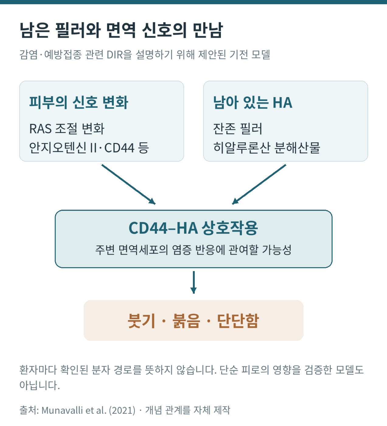
One proposed explanation for reactions following infections or vaccinations involves hyaluronic acid fragments binding to CD44, a receptor found on many cell surfaces. The model proposes that shifts in cutaneous RAS may affect angiotensin II, inflammatory signals and CD44 expression. Residual filler and breakdown products could then play a part in an immune process involving T cells and macrophages, with swelling and firmness. This remains a hypothesis to explain the reported cases. Original mechanistic model
Long-term comfort depends on more than the filler’s name. I consider which tissue receives it, its physical properties and how much is left there. Anatomical research has found differences in how products distribute within tissue, which is why I consider material properties alongside depth and volume. Clinical and anatomical study
4. My approach: address deep support and thin skin separately
In more than 12 years of injecting filler, I have rarely encountered migration or delayed nodules in my own practice. I do not try to solve every under-eye concern with filler. I use a minimal amount and separate the area that needs support from the thin skin that needs attention.
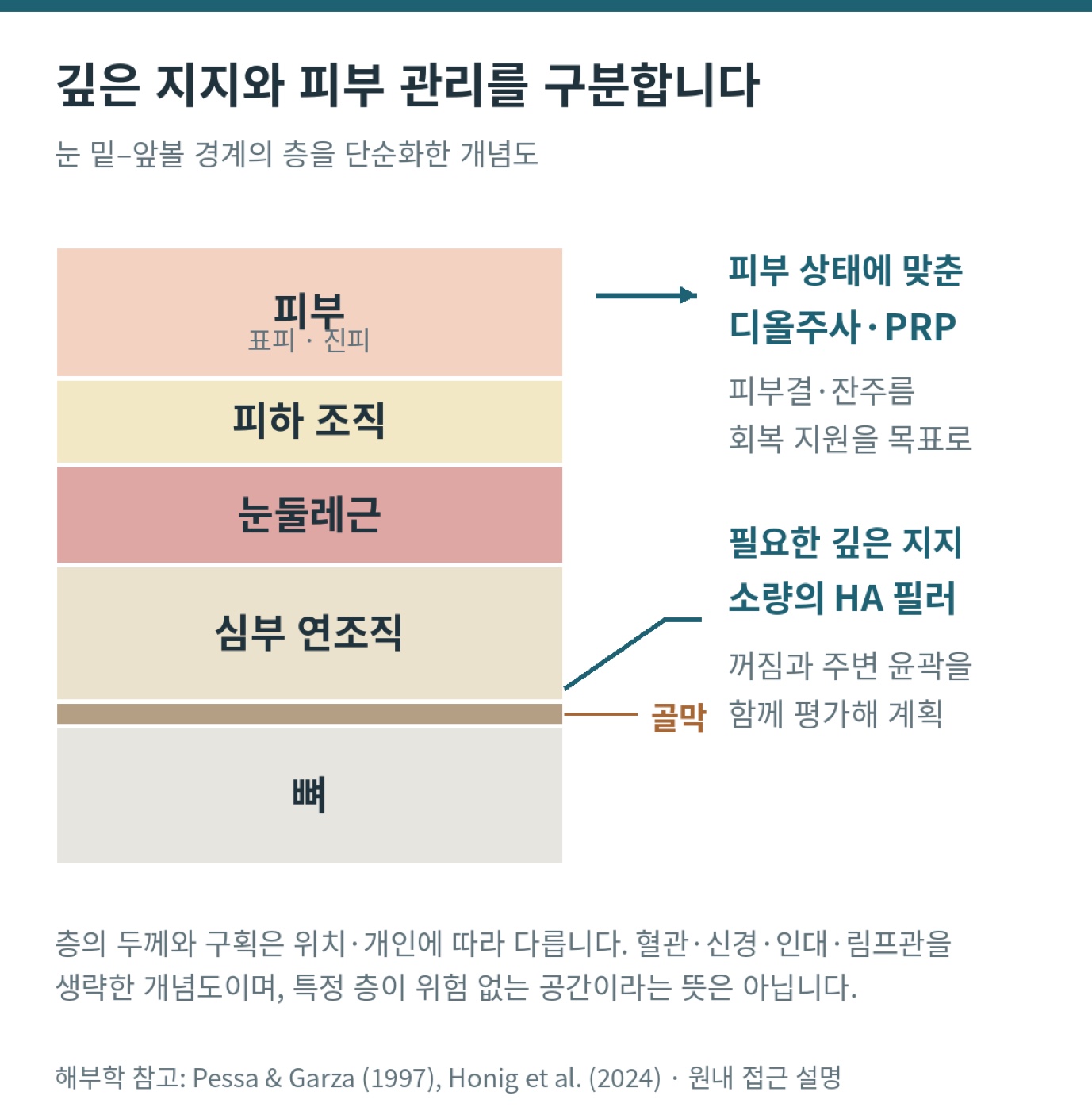
Small-volume structural support for deeper hollowing
If the tear-trough shadow comes from inadequate deep support and filler is appropriate, I may use a small amount of hyaluronic acid in the deep plane above the periosteum of the lower orbital rim. I aim for a smooth transition from the eye to the upper cheek. In my approach, keeping filler largely out of the skin may minimize unnecessary tissue exposure while preserving a natural contour. Heavy-looking under-eye skin can make the middle of the face look weighed down and accentuate the nasolabial folds.
D-ALL and autologous PRP for thin skin
Deep support does not fix thin skin, fine lines or coarse texture. D-ALL is our clinic’s skin-directed injection program, tailored to the patient using components such as PDRN. I sometimes complement it with autologous platelet-rich plasma (PRP) prepared from the patient’s own blood. I avoid products such as Juvelook and Sculptra in this area because of my concerns about inflammatory reactions.
Concentrated platelets in PRP release factors such as PDGF and TGF-β, which participate in tissue repair. In a 2026 split-face study of PRP and PRF around the eyes, 24 people completed the study. Both treatments showed improvements in measures of pigmentation, fine lines and mild hollowing; severe hollowing was not adequately corrected.
Filler and skin-directed treatment can complement each other because under-eye hollowing has multiple causes. Where skin laxity and the collagen framework need attention, I may consider options such as Virtue microneedling radiofrequency or Ultherapy focused ultrasound as part of a tailored plan.
5. If swelling keeps recurring, consider dissolving existing filler
Adding new material over a swollen filler site can make the problem harder to identify. Start by reviewing the treatment history and current symptoms. Ultrasound is useful when needed to locate residual filler and assess surrounding tissue. Depending on the cause, consider dissolving hyaluronic acid filler with hyaluronidase or treating infection or inflammation.
Once the original problem is stable, reassess any remaining hollows or texture concerns. Treat fat protrusion, eye bags, skin laxity or pigmentation according to the underlying cause. Understand what is happening before deciding what, if anything, to add.
A good result still feels right over time
I want the area to look natural when you smile, feel unobtrusive day to day and fit the rest of the face. That is why I plan only the support a deep hollow needs and tailor care to the thin skin. Small amounts above the periosteum, D-ALL, PRP, radiofrequency and ultrasound serve different purposes within that plan.
References and notes
- Bakay ÖSK, Gül B, Kaçar NG. Delayed Inflammatory Reaction to Periorbital Hyaluronic Acid Filler: Relapse After Lisinopril Withdrawal and Response to Retreatment. Journal of Cosmetic Dermatology. 2026;25(11):e71281.
- Munavalli GG, et al. Oral angiotensin-converting enzyme inhibitors for treatment of delayed inflammatory reaction to dermal hyaluronic acid fillers following COVID-19 vaccination—a model for inhibition of angiotensin II–induced cutaneous inflammation. JAAD Case Reports. 2021;10:63–68.
- Pessa JE, Garza JR. The malar septum: the anatomic basis of malar mounds and malar edema. Aesthetic Surgery Journal. 1997;17(1):11–17.
- Funt DK. Avoiding Malar Edema During Midface/Cheek Augmentation with Dermal Fillers. Journal of Clinical and Aesthetic Dermatology. 2011;4(12):32–36.
- Honig SE, et al. Multimodal Pilot Evaluation of a Hyaluronic Acid Infraorbital Filler Using Precise, Multipositional, 3-Dimensional Imaging Quantification, Patient-Reported Outcomes, and Anatomic Cadaveric Assessments. Aesthetic Surgery Journal Open Forum. 2024;6:ojae086.
- Lee YJ, et al. Comparison of the effects of polynucleotide and hyaluronic acid fillers on periocular rejuvenation: a randomized, double-blind, split-face trial. Journal of Dermatological Treatment. 2022;33(1):254–260. (online 2020).
- Behrangi E, et al. Comparing the Efficacy and Safety of PRP and PRF on Periorbital Skin Quality: A Randomized Split-Face Clinical Trial. Journal of Cosmetic Dermatology. 2026;25(6):e70971.
- FDA. Dermal Fillers (Soft Tissue Fillers).