Sometimes skin looks smoother, yet the cheeks and the area around the mouth still seem to sag. After a lifting treatment, the outline may look slimmer, but hollowing in the cheeks can leave the face looking more tired.

Why does this happen?
When I assess facial aging, I do not think we can look at the skin alone. The overall impression comes from skin elasticity, the distribution of fat, and support from bone and ligaments working together.
One area attracting attention in skin biology is dermal white adipose tissue, or dWAT. Fat close to the skin provides volume, but it also exchanges signals with surrounding cells and participates in the environment that helps skin maintain and repair itself.
From this perspective, restoring a youthful face means thinking about what to preserve and restore, as well as how much to tighten.
Fat near the skin talks to the cells that make collagen
The dermis contains fibroblasts, which produce collagen and other components of the extracellular matrix. Think of them as the cells that build and maintain the mesh supporting the skin.
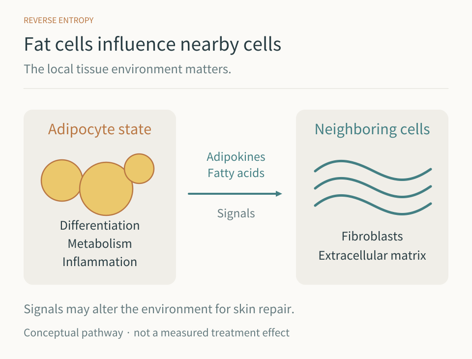
They do not work alone. They adjust their activity in response to signals from immune cells, blood vessels, and nearby fat cells. Fat cells release signaling molecules such as adiponectin, as well as metabolic substances including fatty acids.
When the condition of fat cells changes, the environment in which neighboring fibroblasts work can change too.
A 2021 study of adipocyte–fibroblast metabolism showed that changes in fat cells can influence the production of extracellular matrix by fibroblasts. In other words, adipose tissue is closely connected to the skin’s capacity for renewal.
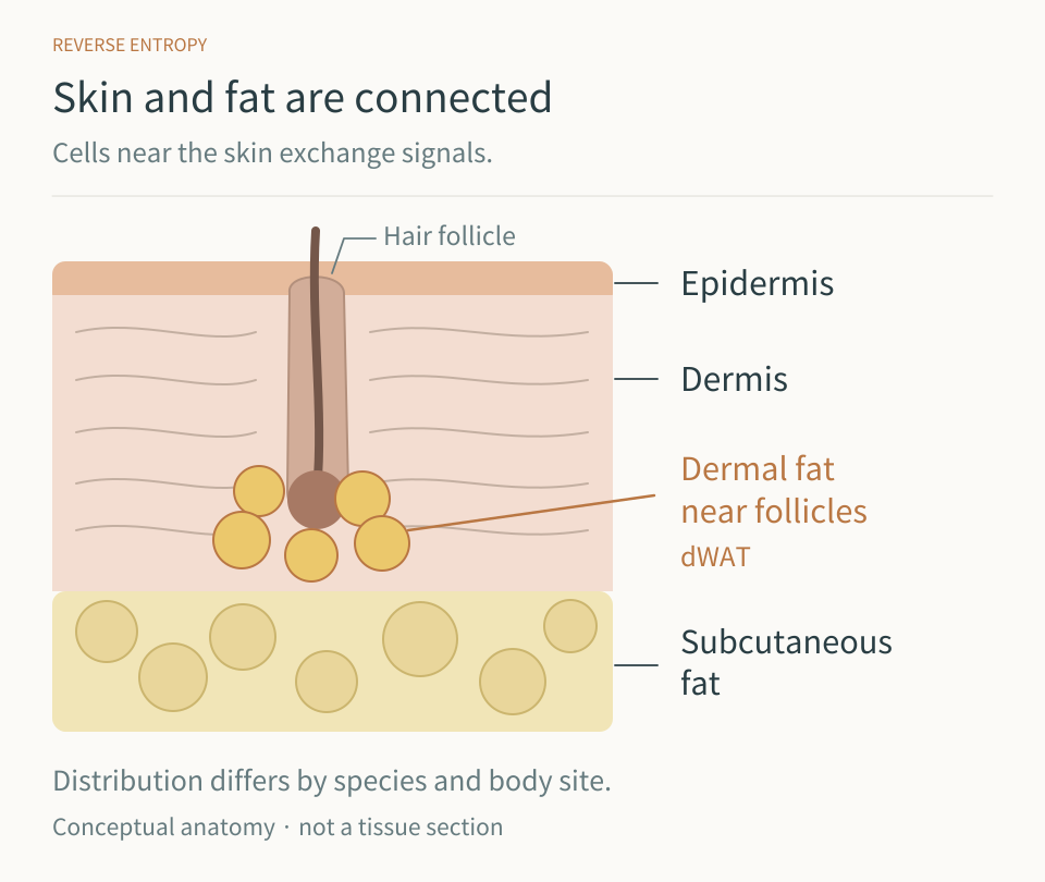
I see these studies as broadening the goals of skin treatment. We can stimulate the cells that make collagen while also considering the surrounding conditions that allow those cells to keep working well.
Aging changes both the amount of fat and how it functions
Facial fat does not decrease uniformly with age. Some areas become hollow, others look relatively prominent, and the structures supporting the tissues also change.
Changes occur at the cellular level too. The ability of fat cells to differentiate and carry out normal metabolism interacts with local inflammation and tissue fibrosis. Aging, ultraviolet exposure, and hormonal changes may all contribute.
Adiponectin is a particularly interesting signal. It is involved in the regulation of metabolism and inflammation in adipose tissue, and researchers are examining its relationship with fibroblasts and maintenance of the skin’s extracellular matrix. Preserving healthy adipose tissue may therefore also help protect the environment in which skin renews itself.
I follow this research closely. We still need to understand how much each of these changes matters in the human face. Even so, the biology helps explain why we should be cautious about indiscriminately reducing fat beneath the skin.
With hollow cheeks, there are questions to ask before choosing stronger lifting
A face that looks as though it is sagging does not always need stronger skin contraction.
Shadows caused by loss of volume in the front of the cheeks, hollow temples, and loose skin can all create a tired appearance. But each calls for a different approach.
If a face already lacks volume, additional fat damage can worsen its three-dimensional shape even when the skin becomes tighter. This is why I pay close attention to fat distribution and thickness before an energy-based procedure.
With both ultrasound and radiofrequency, the depth and amount of energy delivered matter. The same device can produce different results depending on the target tissue and settings. Treatment planning begins by distinguishing tissue that should be reduced from tissue that should be preserved.
My emphasis is on preserving the fat that is needed while encouraging renewal within the dermis. Research on dWAT helps me understand the biology behind that clinical judgment.
What do I consider when planning treatment?
First, deep support and facial volume. When aging changes the shape of bone and deep fat, the contours of the tissues above them also change. Where appropriate, hyaluronic acid filler can be considered to supplement lost volume and support. The hollow areas, treatment depth, and amount must be assessed first. Placement just above the periosteum can help support the overlying tissues and address sagging.
Second, stimulation that encourages dermal renewal. When using radiofrequency, I consider skin thickness, the treatment area, and the fat that needs to be preserved, rather than simply increasing intensity. The aim is to deliver heat where collagen remodeling is needed, within a range the tissue can recover from. I mainly use microneedle radiofrequency, which allows the treatment depth to be adjusted precisely.
Third, the conditions in which stimulated tissue recovers. This is also why I consider treatments such as polynucleotides (PN), represented by products such as Rejuran, and polydeoxyribonucleotide (PDRN) products, often called “salmon injections” in Korea. These substances differ in molecular characteristics and evidence, but both are being studied in relation to tissue repair and skin condition. In my practice, I use these products as part of supporting recovery after energy-based treatments.
What I hope to support is the function of both the dermis and the tissues beneath it. How far such combinations can improve the signaling environment of dWAT is something I would like future research to clarify. I am bringing adipose-tissue biology into treatment planning alongside established principles of anatomical support and skin renewal.
Deep support
Assess volume loss and facial shape.
Supplement structural support when appropriate.
Dermal renewal
Match energy delivery to skin condition.
Plan stimulation within the tissue’s capacity to recover.
Fat and recovery
Preserve the fat that is needed.
Consider the environment for repair after treatment.
I think about the face beyond tighter skin
Tighter skin and a younger-looking face are not always the same thing. Natural volume, soft curves, skin quality, and expression need to work together.
That is why I try to connect the skin’s surface condition, dermal elasticity, the distribution and function of fat, and deeper structural support, rather than ending the assessment at the skin.
Understanding the role of each layer matters.
As research on dWAT and other tissues around the skin develops, we should gain a clearer understanding of which fat to preserve and which conditions for repair to support.
I hope facial rejuvenation continues to move toward preserving good structure and the capacity to recover over time, rather than concentrating only on stronger lifting.

References
- Zhang et al. Dermal adipocytes contribute to the metabolic regulation of dermal fibroblasts. Experimental Dermatology. 2021. Article.
- Menopause and Dermal White Adipose Tissue Depletion: Mechanistic Links, Adipogenesis, and Regenerative Therapeutic Replacement. Journal of Cosmetic Dermatology. 2026. Article.
- Widgerow AD. Adipose Tissue, Regeneration, and Skin Health: The Next Regenerative Frontier. Aesthetic Surgery Journal Open Forum. 2024. Article.
- Rho et al. Expert Perspectives: Evidence-Based Applications of Polynucleotides (PNs) in Aesthetic Medicine and Dermatology. 2026. Article.